Mata Chanan Devi Hospital C-1, Janakpuri, New Delhi -110058
 + (91) - (11) - 45582000/10
Mata Chanan Devi Hospital C-1, Janakpuri, New Delhi -110058
+ (91) - (11) - 45582000/10
The Department of Radiology and Imaging services is located on the ground floor of the new building of hospital. The Radiology & Imaging at MCDH is a highly advanced facility that is equipped with the latest advanced state-of-the-art technology. The department is self contained with a separate reception, waiting area, examination and equipment room.
It is a technique used in Radiology to visualize in detail the internal structures of the body. It provides excellent contrast between the various soft tissues of the body. It is very useful in Imaging Brain, Spine, Joints, Muscles & Pelvic Organs including Prostate and Ovaries. Compared with other medical Imaging techniques, it does not use X-Ray radiation. It is safe in pregnancy and newborn children.
We have the State-of-the-art Siemens MRI machine. Specialized MR studies like Post Gadolinium Contrast Studies, MRI Brain with Epilepsy Protocol, MRCP, Angiography and Venography are also done.
Whole Spine Screening is possible in patients undergoing MR for disc prolapse, Sciatica, back pain and cancers.
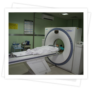
Computed Axial Tomography (CAT) is an imaging method using focused X-ray beam to generate an image of a body part in an axial plane to acquire a 3-D data.
All CT scan studies are performed including specialized procedures like CT angiography(Cerebral , Peripheral, Pulmonary and Abdominal Angiographies), Liver, Kidney, Oncology Imaging , Non Invasive CT Bronchoscopy and Colonoscopy, 3D Reconstruction of Joints and Musculoskeletal System in Tumors and Trauma, CT guided FNAC’S, biopsies and Drain placements are done in routine practice.
Each patient is evaluated for safety of Contrast Administration and providing Anaesthesia / Sedation whenever required. There are extensive discussions with the referring clinicans before and after the scans and all scans are interpreted by highly qualified, well trained, experienced Radiologists in view of the clinical findings. The Department meets all AERB guidelines for Radiation practice, running teaching programs and is doing research in various subspecialties of radiology.
Seven Ultrasound Machines with Colour Doppler equipped with transvaginal, transrectal, linear & cardiac probes
Ultrasound facility is available round the clock thus facilitating immediate life saving intervention in emergency situations such as trauma cases, appendicitis, ectopic pregnancies, perforations, etc.
USG machine is taken for bedside sonography of sick patients and guided procedures in O.T. & ICU.
The radiology department is equipped with a number of conventional x-ray machines and One machine with image intensifier for procedures to be done under fluoroscopic control. The department work load on an average is 120 patients per day. Of this, the bulk of the work comprises of routine radiological examination of chest, abdomen, spine and extremities.
The Radiology Department has been running a post-graduate training course the National Board of Examinations (D.N.B. degree in radiology) since 2012. The department imparts training in both conventional radiology and modern imaging techniques of ultrasound, CT and MRI. A well planned teaching and training programme, spread over three years is being conducted so as to make the students fully competent to practice ,teach & do research in various subspecialities of radiology